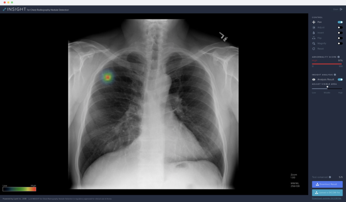
The technology that can empower medical treatment and predict problems, especially lung cancer, tuberculosis, pneumonia, and pneumothorax – the four major chest diseases, much more accurately than medical practitioners is being developed by Korean technology experts using Artificial Intelligence (AI).
A joint research team consisting of Park Chang-min, a professor of imaging at Seoul National University Hospital and Lunit, a software development company, announced on April 1, that the accuracy rate exceeded 98 percent after applying the Artificial Intelligence auxiliary diagnosis system to diagnose four major chest diseases. The research team explained that the system not only detected lung diseases but also found location.
The assessment of the diagnosis was conducted at Seoul National University Hospital, Boramae Hospital, Kangdong Kyung Hee University Hospital, Eulji University Hospital and Grenoble University Hospital in France. In the assessment, Artificial Intelligence scored over 97 percent on average. In addition, the accuracy of Artificial Intelligence recorded 98.3 percent in a comparative evaluation with 15 doctors, which is higher than 93.2 percent of the pre-scientists in chest imaging. In medical staff, the reading ability was also increased by up to 9 percentage points when assisted by Artificial Intelligence.
To develop the Artificial Intelligence system, the research team explained that it used the results of 98,621 chest X-ray imaging data related to the four major chest diseases to learn how to diagnose Artificial Intelligence. This is the same principle that AlphaGo learned about Go.
Early treatment through accurate diagnosis is very important because the four major chest diseases have high frequency and mortality rates worldwide. The system is expected to be used to diagnose actual patients after approval of medical devices by the Ministry of Food and Drug Safety . “The Artificial Intelligence system analyzes X-ray images of patients’ chests and indicates areas where abnormalities are found, and presents the possibility as a probability value,” the research team said. “This will make it easier to diagnose images.”


